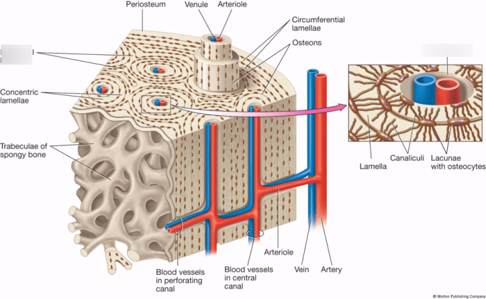
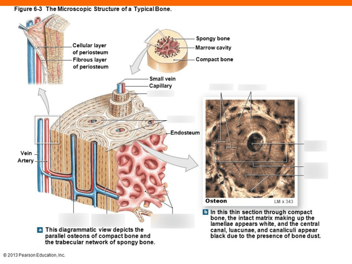
Drag the labels to identify the microscopic structures of bone is an interactive learning tool that provides a comprehensive overview of the intricate components that make up bone tissue. This engaging resource allows users to explore the histological appearance of bone under a microscope, gaining a deeper understanding of the cellular and extracellular components that contribute to its unique properties.
Through a series of interactive exercises, learners can identify and label osteocytes, osteoblasts, and osteoclasts, the key cellular players in bone remodeling. They will also delve into the composition and organization of the organic and inorganic components of bone matrix, including collagen and hydroxyapatite, and discover their roles in bone strength and flexibility.
Bone Structure

Bone tissue is a hierarchical composite material with a complex structure that provides strength, flexibility, and support to the body. It is organized into three levels:
- Macroscopic: The gross anatomy of bone, including its shape, size, and external features.
- Microscopic: The cellular and extracellular components of bone, including osteocytes, osteoblasts, osteoclasts, and the bone matrix.
- Nano: The molecular structure of bone, including the arrangement of collagen fibers and hydroxyapatite crystals.
Cortical and Trabecular Bone
Bone can be classified into two main types based on its structure and function:
| Characteristic | Cortical Bone | Trabecular Bone |
|---|---|---|
| Structure | Dense, solid outer layer | Porous, cancellous inner layer |
| Function | Provides strength and support | Absorbs shock and reduces stress |
| Location | Outer surface of long bones, shaft of long bones | Inner core of long bones, vertebrae, pelvis |
Microscopic Structures of Bone

Under a microscope, bone tissue exhibits a distinct cellular and extracellular architecture.
Cellular Components
- Osteocytes: Mature bone cells that maintain the bone matrix and sense mechanical stress.
- Osteoblasts: Bone-forming cells that secrete the organic components of the bone matrix.
- Osteoclasts: Bone-resorbing cells that break down the bone matrix to release calcium and phosphate ions.
Diagram of Cellular Components of Bone
[Diagram yang menunjukkan osteosit, osteoblas, dan osteoklas yang mengelilingi matriks tulang]
Bone Matrix

The bone matrix is composed of an organic component (mainly collagen) and an inorganic component (mainly hydroxyapatite).
Organic Component, Drag the labels to identify the microscopic structures of bone
Collagen is the main protein in bone and provides flexibility and tensile strength.
Inorganic Component
Hydroxyapatite is a calcium phosphate mineral that gives bone its hardness and compressive strength.
Bone Mineralization
Bone mineralization is the process by which hydroxyapatite crystals are deposited in the collagen matrix, giving bone its rigidity.
Bone Remodeling: Drag The Labels To Identify The Microscopic Structures Of Bone

Bone remodeling is a continuous process that involves the breakdown of old bone and the formation of new bone to maintain bone health and strength.
Cellular and Molecular Mechanisms
- Osteoclastsbreak down bone matrix, releasing calcium and phosphate ions.
- Osteoblastsform new bone matrix, using the released calcium and phosphate ions.
- Hormones(e.g., parathyroid hormone, calcitonin) and mechanical stressregulate bone remodeling.
Clinical Implications of Impaired Bone Remodeling
Impaired bone remodeling can lead to bone diseases such as osteoporosis and Paget’s disease.
Bone Diseases
| Disease | Causes | Treatments |
|---|---|---|
| Osteoporosis | Reduced bone density and strength | Calcium supplements, bisphosphonates, hormone replacement therapy |
| Paget’s Disease | Abnormal bone remodeling | Bisphosphonates, calcitonin |
| Osteomalacia | Deficiency of vitamin D or calcium | Vitamin D supplements, calcium supplements |
Imaging Techniques in Diagnosing Bone Diseases
X-rays, CT scans, and MRI scans are used to diagnose bone diseases by visualizing bone structure and density.
General Inquiries
What are the advantages of using drag the labels to identify the microscopic structures of bone?
Drag the labels to identify the microscopic structures of bone offers several advantages, including:
- Interactive and engaging learning experience
- Enhanced visualization and comprehension of bone structures
- Self-paced and personalized learning
- Immediate feedback and reinforcement of understanding
What types of bone cells can be identified using drag the labels to identify the microscopic structures of bone?
Drag the labels to identify the microscopic structures of bone allows users to identify three main types of bone cells:
- Osteocytes: Mature bone cells that maintain bone tissue
- Osteoblasts: Bone-forming cells that secrete new bone matrix
- Osteoclasts: Bone-resorbing cells that break down old bone tissue
How does drag the labels to identify the microscopic structures of bone contribute to a deeper understanding of bone biology?
By actively labeling and identifying the microscopic structures of bone, learners gain a deeper understanding of:
- The cellular and molecular components of bone tissue
- The processes of bone formation, remodeling, and repair
- The impact of various factors on bone health and disease